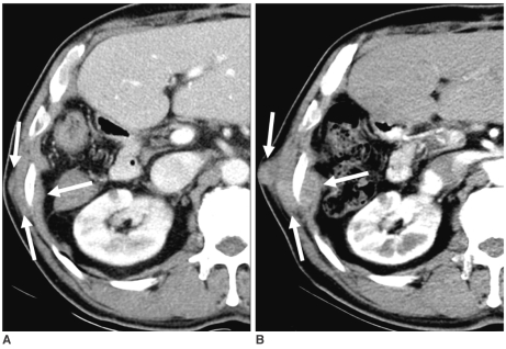
Fig. 3.
66-year-old woman with needle tract implantation with large conglomeration around rib after percutaneous hepatocellular carcinoma biopsy, followed by right hepatic lobectomy.
A. Contrast-enhanced CT image showing isoattenuation nodules (arrows) relative to intercostal muscles in intercostal muscles and subcutaneous fat layer five months after percutaneous biopsy using end-cutting needle.
B. Follow-up CT image showing conglomerated nodules (arrows) with increased size nine months after percutaneous biopsy.

