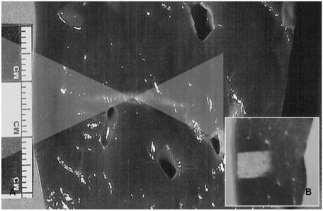
Fig. 5.
Classical thermal lesion formed by focused US surgery (US absorption only) on porcine liver specimen.
A. Cigar-shaped thermal lesion is formed at focal zone of US wave pathway (two overlaid triangles) following HIFU single exposure.
B. Final thermal lesion after stacking each single lesion. Single lesions are much smaller than clinically common tumors and therefore each thermal lesion should be stacked compactly without leaving intervening viable tissue. This lesion can cover entire pathological lesion as well as has very sharp margin that could be controlled easily.

