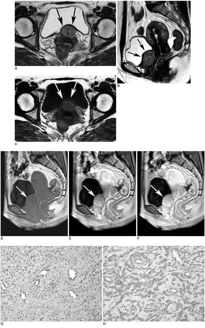
Fig. 1.
Angiomyofibroblastoma in 48-year-old woman.
A, B. T2-weighted axial (A) and sagittal (B) MR images show well-defined, oval shape mass with heterogeneous signal intensity in posterior perivesical space (arrows). Note nodular or curvilinear dark signal intensities within tumor.
C. On T1-weighted axial MR image (10-minutes after contrast administration) at same level as in A, mass (arrows) is isointense to muscle.
D-F. Contrast-enhanced dynamic sagittal MR images (D: baseline, E: 1-minute, F: 3-minute delayed image) show strong, homogeneous and persistent enhancement of mass (arrows).
G. Photomicrograph of surgical specimen shows compactly arranged epithelioid ovoid or blunt spindle shaped tumor cells having monotonous small nuclei and eosinophilic cytoplasm. There are prominent ectatic blood vessels, which are surrounded by eosinophilic or myxoid fibrous stroma (Hematoxylin & Eosin staining, original magnification, ×200).
H. Immunohistochemical staining shows that neoplastic cells are strongly positive (brown color) for actin (original magnification, ×200).

