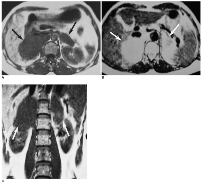
Fig. 1.
Abdominal MRI demonstrates that the masses in the bilateral adrenal glands are homogeneous hypointense to the liver (black arrow) on the axial T1-weighted spin echo image (A) and hyperintense (white arrow) on the axial T2-weight spin echo image (B). The mass in the right adrenal region has invaded the inferior vein cava (A, B) and upper polar of the right kidney (white arrow) on the coronal T1-weighted spin echo image (C).

