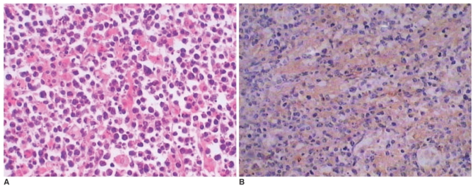
Fig. 2.
A. Photomicrography of the histology specimen shows the tumor consists of poorly-differentiated plasma cells with large and hyperchromatic nuclei (Haematoxylin & Eosin, × 300).
B. Immunohistochemical staining of the resected masses is positive for the monoclonality of IgG-lambda (Immunohistochemical staining, × 200).

