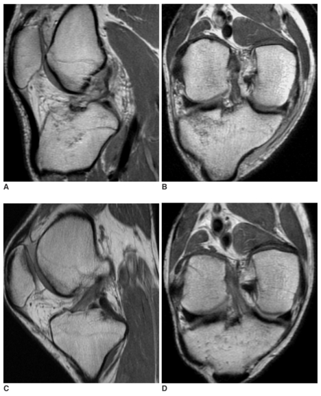
Fig. 1.
A 25-year-old man with a completely healed anterior cruciate ligament that was confirmed by arthroscopy.
A, B. Oblique sagittal (A) and coronal (B) proton density-weighted images obtained on the day of the knee trauma show complete tear at the proximal anterior cruciate ligament and associated fracture at the lateral tibial plateau.
C, D. Follow up oblique sagittal (C) and coronal (D) images after four-months reveal a nearly normal-looking anterior cruciate ligament. The anterior cruciate ligament appears to show a low signal intensityspotted with a high signal intensity, a sharp contour, a straight course and a normal thickness. Arthroscopy performed 2-months later, due to contracture of the knee joint by the hypertrophy of ligamentum mucosum, demonstrated a normal appearance with normal tension of the anterior cruciate ligament. The side-toside difference of the KT-2000 measurement was 3 mm at the time of arthroscopy.

