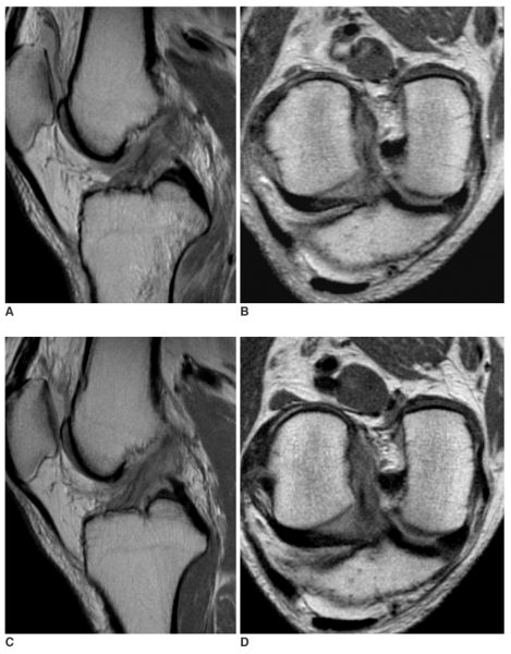
Fig. 2.
A 54-year-old man with partial tear of anterior cruciate ligament that was confirmed by arthroscopy.
A, B. Sagittal (A) and oblique coronal (B) proton density-weighted images 15 days after the trauma show complete tear at the proximal anterior cruciate ligament and marrow contusion at the posterior aspect of the proximal tibia.
C, D. Four-months later, the sagittal (C) and oblique coronal (D) images demonstrate restored continuity of anterior cruciate ligament, which shows a band-like high signal intensity within the ligament, a partly obscure contour, mild sagging and an increased thickness. Partial tear of the anterior cruciate ligament was confirmed during arthroscopy 5-months later. The side-to-side difference of the KT-2000 measurement was 5 mm at the time of arthroscopy.

