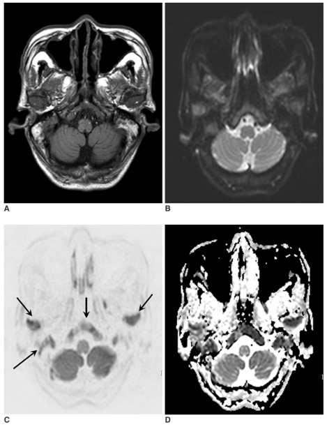
Fig. 2.
A 69-year-old man with lung cancer (case no. 3). T1-weighted image (A), B0 image (B), isotropic diffusion-weighted image (C), and an apparent diffusion-coefficient map (D) were obtained. Lesions involving the skull base and mandibular condyles (arrows) are visible on all images. However, the isotropic diffusion-weighted image shows a superior lesion contrast.

