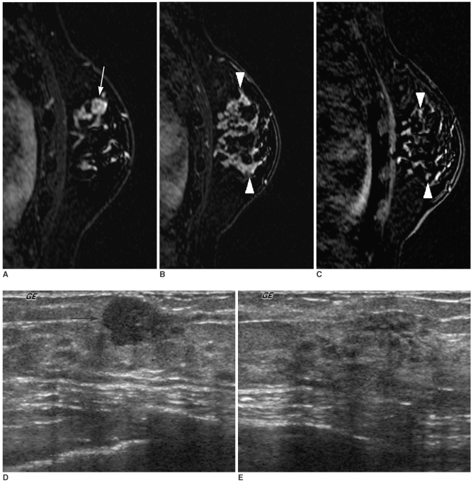
Fig. 3.
A 40-year-old woman with a 1 cm palpable cancer of the left breast.
A, B. Dynamic enhanced and subtracted T1-weighted sagittal MR images show a 1 cm intensely enhancing main mass (arrow) (A) in addition to a 5 cm clumped segmental enhancement with washout (arrowheads) (B) in a different plane.
C. A high signal intensity (arrowheads) of a 5 cm clumped segmental enhancement is noted on the reverse subtracted image.
D. Initial US shows only a 1 cm hypoechoic mass (arrow) at a palpable site.
E. Targeted US shows a normal-looking, but slightly heterogeneous parenchyma. We interpreted that the additional enhancing lesion on MR had no US correlate. On a frozen section examination during the surgical operation, the additional enhancing lesion at the surrounding parenchyma revealed a malignancy, which was confirmed as an extensive intraductal carcinoma with an invasive ductal carcinoma following mastectomy. The surgical plan was changed from conservative breast surgery to mastectomy without the consent of the patient.

