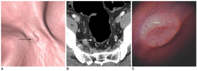
Fig. 1.
An 8 mm flat-elevated rectal lesion in a 50-year-old man.
A. A three-dimensional lumen view of the proximal rectum revealing an 8 mm, flat-elevated lesion with central depression (arrow) on CT colonography.
B. A contrast-enhanced axial CT image at the level of the proximal rectum shows a small enhancing flat-elevated lesion (arrow).
C. A photograph from conventional colonoscopy displays a flat-elevated lesion with central depression in the proximal rectum. Histological examination revealed that the lesion was an adenocarcinoma.

