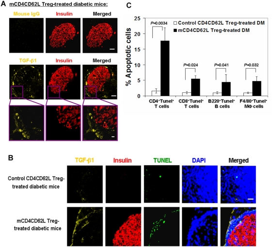
Figure 4. Treatment with mCD4CD62L Tregs enhance expression of TGF-β1 in pancreatic islets.
Overt diabetic NOD mice treated with mCD4CD62L Tregs were sacrificed for pancreatic immunohistochemistry studies after observation for 45 days (n = 8). Representative data are from 6 diabetic mice (6/8 mice) sensitive to mCD4CD62L Treg treatment with euglycemia. The control CD4CD62L Treg-treated diabetic mice served as control (n = 5). (A) TGF-β1 staining surrounds a pancreatic islet of mCD4CD62L Treg-treated diabetic mice, as determined by double-immunostaining for TGF-β1 (yellow) and insulin (red). TGF-β1 positive cells (bright yellow) and released TGF-β1 in matrix (faint yellow) were distributed in the islet β cell area (red) and surrounded islet β cells (top panels, scale bar 50 µm). High magnification is shown in bottom panels, scale bar 10 µm. Isotype-matched mouse IgG1 served as a negative control for TGF-β1 immunostaining in a serial pancreatic section. Representative images were obtained from five experiments. (B) TUNEL assay. The proteinase K-pretreated pancreatic slides were initially immunostained with TGF-β1 (yellow) and insulin (red) Abs, followed by TUNEL assay (green), and nuclear counterstaining with DAPI (blue). Scale bar 20 µm. Representative images were obtained from four experiments. (C) Percentage of apoptotic cells in subtypes of infiltrated leukocytes in pancreatic islets. Cryosections (8 µm thickness) of frozen pancreata from mCD4CD62L Treg-treated diabetic mice (n = 4) and control mice (n = 4) were initially detected with In Situ Cell Death Detection Kit (Roche), followed by immunostaining with different monoclonal Abs and imaging with a Zeiss LSM 510 META confocal microscope. Cryosections incubated with label solution without the TUNEL reaction mixture and/or isotype-matched IgG served as negative controls. Data represent mean±s.d. of five experiments.

