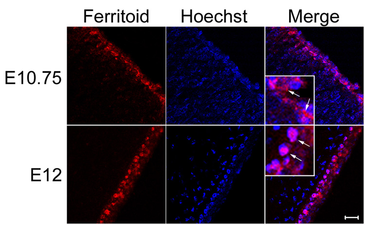
Figure 4. Confocal micrographs of ferritoid at stages before the appearance of ferritin (E10.75), and after the appearance of ferritin (E12).
When these images are merged with that of Hoechst staining, it can be seen that at E10.75 the ferritoid in CE cells is cytoplasmic and at E12 it is nuclear (the inserts are enlarged images in which the arrows point to cytoplasmic staining at E10.75 and nuclear staining at E12). Scale bar in bottom right panel = 10 µm.

