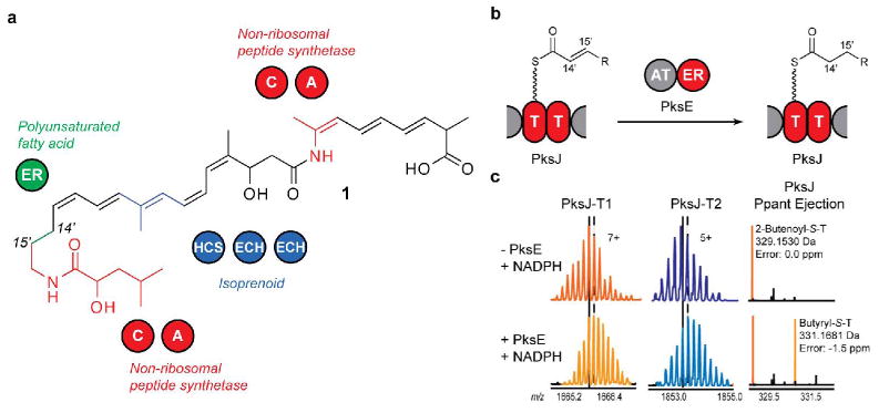
Figure 1.

(a) Structure of dihydrobacillaene. C, condensation; A, adenylation; HCS, HMG-CoA synthase; ECH, enoyl-CoA hydratase. (b) PksE reduces an enoyl substrate tethered to the PksJ T-domain doublet in trans. (c) Confirmation of PksE enoyl reductase activity. (Left) PksJ-T1 active-site peptide. (Middle) PksJ-T2 active-site peptide. (Right) Ppant ejection ions. Vertical lines in (c) correspond to the most abundant isotopic peak (solid: absence of PksE; dash: presence of PksE).
