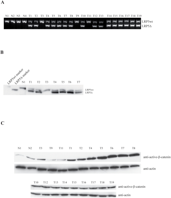
Figure 1. The LRP5Δ receptor is expressed in breast tumors.
Accumulation of active β-catenin. (A) PCR analysis of cDNA from normal breast tissue (N1–N4) and primary breast cancer (T1–T19). Primers were located in exons 9 and 13 of LRP5 as described [26]. The PCR reaction is not quantitative as the primers compete for the two fragments. A total of sixty-two LRP5 truncated fragments (see also Table 1) were directly sequenced and all contained the same in-frame deletion of 142 amino acids (Δ666–809). (B) Immunoprecipitation and Western blot analysis of LRP5. Tissue numbering corresponds to panel A. Transiently expressed LRP5wt and LRP5Δ shown as markers. (C) Western blot analysis of non-phosphorylated active β-catenin. Tissue numbering as in panel A.

