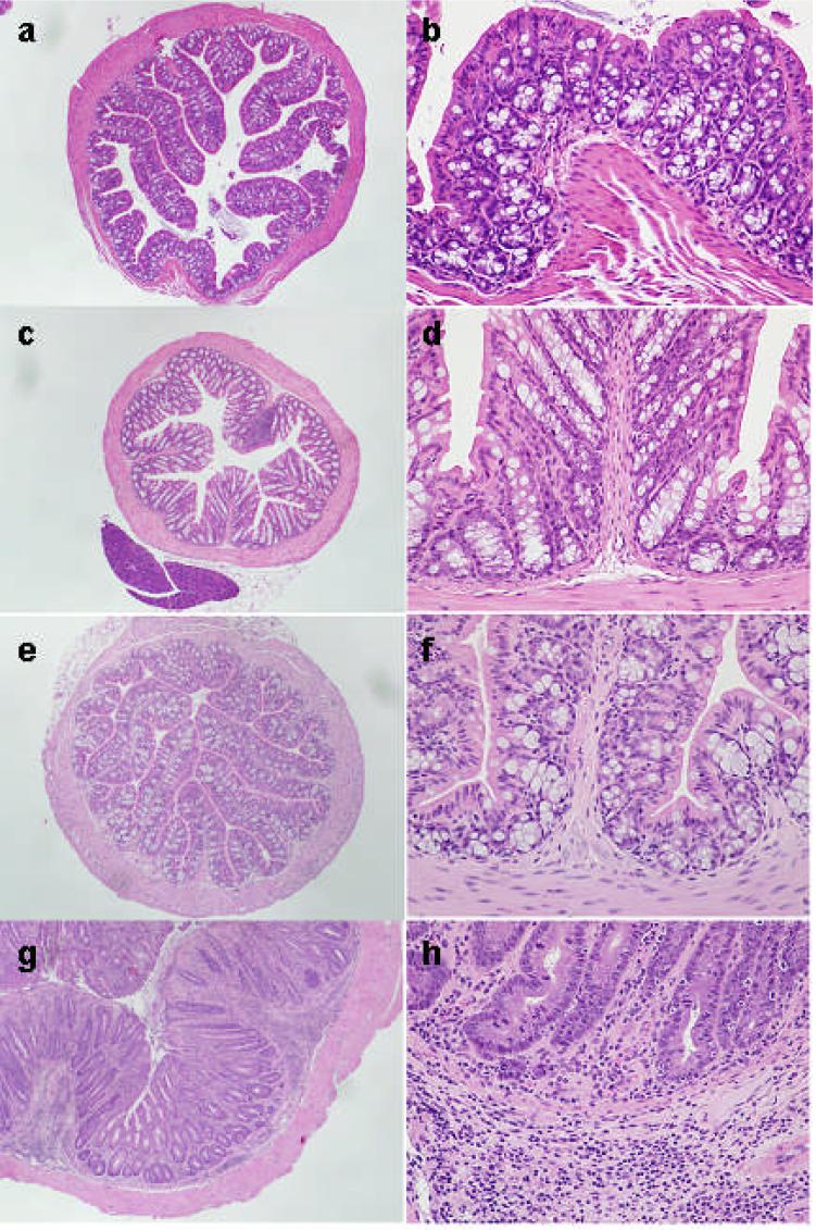
Figure 1.
H&E stained sections of colons from 4 month old mice. (a,b) wild-type C57BL/6 (B6) mouse, (c,d) Gαi2-deficient B6 mouse, (e,f) wild-type 129SvEv (129) mouse and (g,h) Gαi2-deficient 129 mouse. (a) through (f) show show overall preserved mucosal architecture, cytology and mucin production. A few lymphocytes are present in the lamina propria. Epithelial cells do not show increased mitotic activity or reactive changes. The Gαi2-deficient 129 mouse colon displays architectural distortion with occasional bifurcation of glands, mucosal mucin depletion and predominantly plasma lymphocytic infiltrate, involving the lamina propria, muscularis mucosa and submucosa. Inflammation also contains rare eosinophils and occasional neutrophils. There is focally prominent mitotic activity in the glandular epithelium. Epithelial cells show reactive changes, with hyperchromasia and enlargement of nuclei, irregularity of the nuclear contours and occasionally prominent nucleoli. No atypical mitoses are seen. Magnification 40x (a,c,e,g) and 200x (b,d,f,h)

