Figure 3.

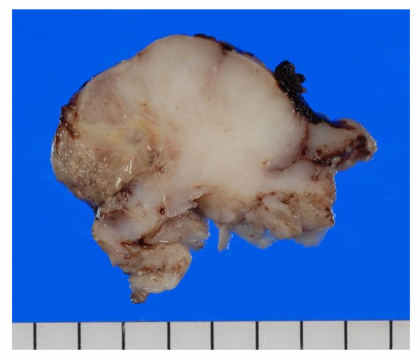
Macroscopic image of the tumor. The lesion was a fragile mass with ulcerated surface. The cut surface was grayish-white in color, myxoid or lobular pattern in some areas.

Macroscopic image of the tumor. The lesion was a fragile mass with ulcerated surface. The cut surface was grayish-white in color, myxoid or lobular pattern in some areas.