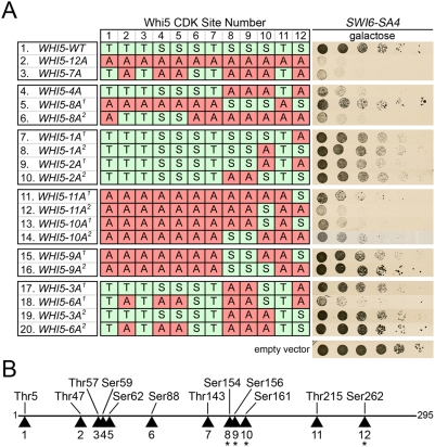
Figure 4. Mutational analysis of Whi5 phosphorylation sites.
(A) swi6Δ [SWI6-SA4] cells expressing Whi5-WT or Whi5 phosphorylation mutants from the GAL1 promoter. Cells were spotted in serial 5 fold dilutions on galactose media and incubated 48 hours. The table indicates the CDK sites of Whi5 that were left wild type (S/T) or mutated to alanine (A) for each mutant construct. (B) Schematic of the Whi5 protein showing relative location of CDK sites, numbered 1–12.

