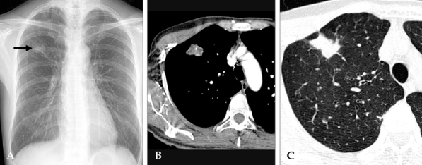
Fig. 1.

45-year-old woman with Mycobacterium avium complex pulmonary disease manifesting as an SPN. (A) The posteroanterior chest radiograph reveals a nodular opacity in the right upper lung zone (arrow). (B) Contrast-enhanced chest computed tomography scan (5-mm collimation) obtained at the level of the aortic arch reveals a solitary peripheral pulmonary nodule located in the right upper lobe. Note the necrotic low-ttenuation portions and tiny calcifications within the nodule. (C) Lung window image reveals the irregular shape of the nodule, which also exhibits some marginal spiculation.
