
Fig. 2.
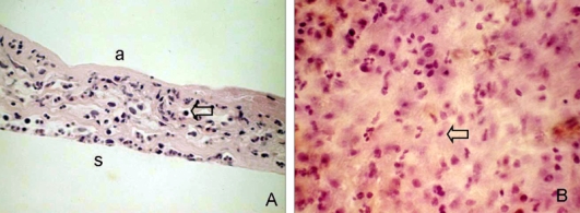
Histological features of a temporary amniotic membrane patch removed 4 days after pterygium excision. Amniotic membrane was sectioned and stained with H&E. (A) In a cross section, one or two layers of epithelium were observed over the partially dissolved amniotic membrane with marked mononuclear cell infiltration (arrow). (B) In a flat mount, numerous round or spindle-shaped mononuclear cells (arrow) had infiltrated and a small number of polymorphonuclear leukocytes were found on the stromal side. a, amnion side; s, amniotic membrane stroma. Original magnification: A 200×; B 400×.