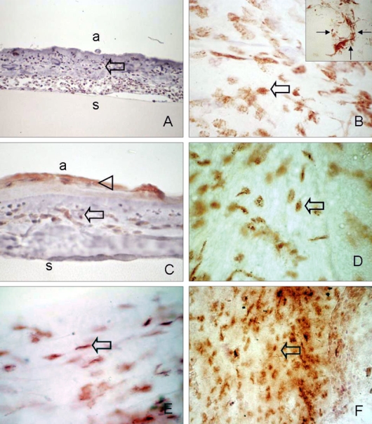
Fig. 3.

Immunohistochemical analysis of infiltrated stem cells in removed amniotic membrane. (A, B) Abundant CD34-positive cells, which were mainly composed of round or spindle-shaped mononuclear cells, were observed on the stromal side of the amniotic membrane (arrow). Occasionally, some CD34-positive cells formed clusters resembling small vessels (B; small arrow, inset). (C, D) c-kit immunoreactivity was found in grown epithelium (arrow head), spindle or round-shaped monocytes on the amniotic membrane (arrow). (E) SRTO-1-positive spindled-shaped cells resembling fibroblasts were observed (arrow). (F) Small round or long slender shaped AC133 positive cells (arrow). Sections were counterstained with hematoxylin. a, amnion side; s, amniotic membrane stroma. Original magnification: A, C 200×; B, B inset, D, E 400×. A, C: cross-section, B, D, E, F: flat mount.
