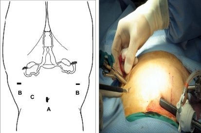
Fig. 3.
Port placement: (A) The 12-mm camera port was placed in the umbilicus or above depending on the size of the uterus. (B) The 8-mm lateral ports for robotic instruments were mounted directly to the robotic arms and placed 2 to 3 cm medial and superior to the anterior superior ileac spine with modification based on the size of the uterus. (C) The assistant port was placed between the camera port and the left lower quadrant port.

