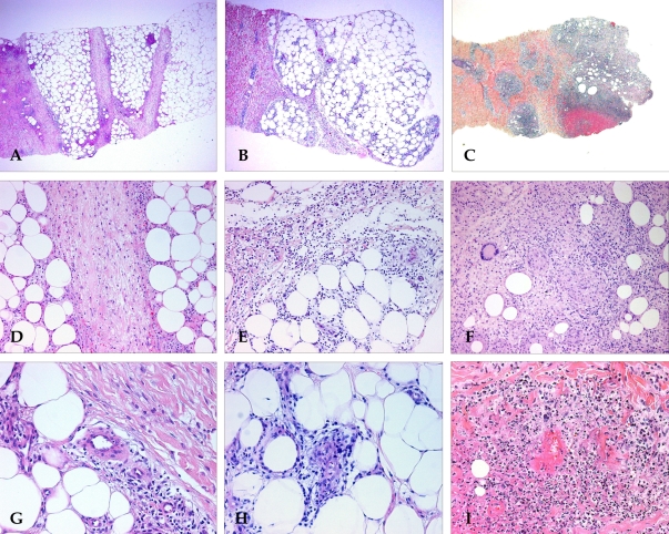
Fig. 2.
Histopathologic findings of EN, EN-like lesion in BD, and NV. Pattern of panniculitis: Mostly septal panniculitis of EN (A, H & E stain, × 40). Inflammation presents mainly in and around the septa. Mixed septal and lobular panniculitis of EN-like lesion in BD (B, H & E stain, × 40). Inflammatory cells diffusely infiltrated in septa and lobules without clear compartmentalization. Mostly lobular panniculitis of NV (C, H & E stain, × 40). A region of caseation necrosis and surrounding inflammation replacing the septa and lobules was observed. Inflammation: There is an edema of the septa with paraseptal inflammation composed of lymphocytes mixed with histiocytes in EN (D, H & E stain, × 200). A dense inflammatory infiltrate of neutrophils mixed with lymphocytes was observed in the EN-like lesion (E, H & E stain, × 200). Severe lobular inflammation in NV (F, H & E stain, × 200), with extensive necrosis of adipocytes and granulomatous inflammation was observed. Vasculitis: No vasculitis was observed in EN (G, H & E stain, × 400). Lymphocytic vasculitis in EN-like lesion (H, H & E stain, × 400). Lymphocytic infiltrate around and within vessel wall was observed. Neutrophilic vasculitis in NV (I, H & E stain, × 400). Destruction of the vascular wall, surrounding neutrophils and nuclear dusts was observed.

