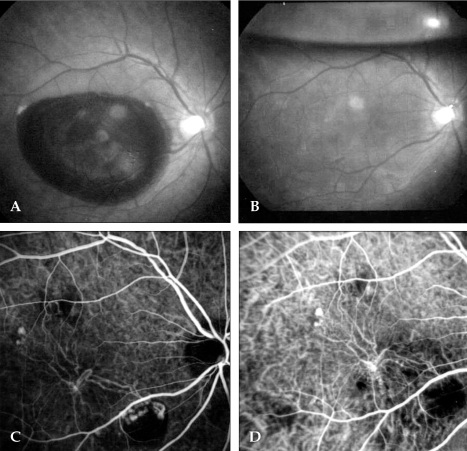
Fig. 3.
PCV in a 55-year-old man. (A) Fundus photo showing thick subretinal hemorrhage in the macula of the right eye. The visual acuity was finger counting. (B) Fundus photo taken one day after tPA and SF6 gas injection, showing absorption of the hemorrhage. The visual acuity was 0.4. (C) Early stage ICG angiogram showing an abnormal choroidal vascular network and polypoid lesions connected to abnormal choroidal vessels inferonasal to the macula. (D) ICG angiogram taken three months after PDT. Polypoid lesions can not be detected and visual acuity was 0.8.

