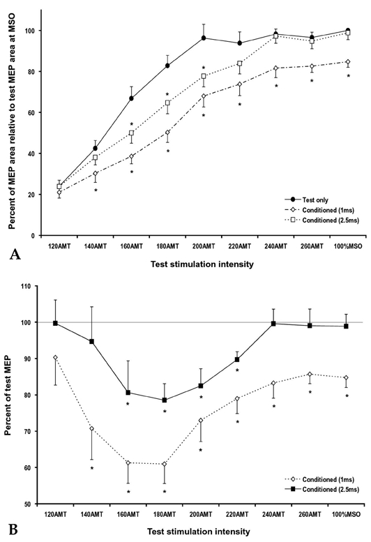
Fig. 1.

(A) Motor evoked potential (MEP) area of the right first dorsal interosseus (FDI) evoked by single- (circle) and paired-pulse TMS with 1 ms (diamond) and 2.5 ms (rectangle) interstimulus intervals (ISIs) at different test stimulation (TS) intensities. MEP areas were significantly suppressed in paired-pulse trials with 1 ms ISI at a TS of 140% or higher of the active motor threshold (AMT) and in paired-pulse trials with 2.5 ms ISI at a TS of 160 - 200% of the AMT as compared with the control trials (*different from the control trials, p < 0.05). (B) Calculated short-interval intracortical inhibition (SICI, % of conditioned MEP area/test MEP area) at different TS intensities. SICI was significant in paired-pulse trials with 1 ms ISI at a TS of 140% or higher of the AMT and in paired-pulse trials with 2.5 ms ISI at a TS of 160 - 220% of the AMT (*p < 0.05).
