
Fig. 2.
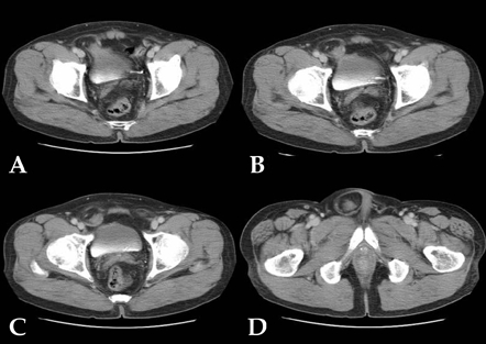
Initial abdomen/pelvis CT showed that the right anteroinferior wall of bladder was herniated into the right inguinal canal without contrast enhancement within the herniated sac. The neck of the hernial sac was relatively wide (A and B) and within the hernial sac there was homogeneous cystic low density due to trapped urine without calcification (C and D).