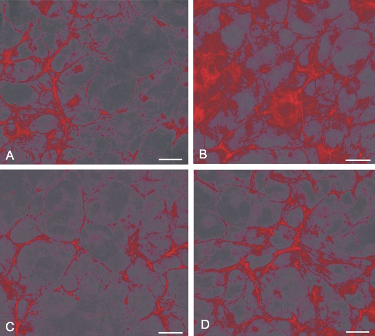
Fig. 3.

Comparative fluorescent micrographs showing IGF-1 inhibition of TGF-β-mediated fibronectin accumulation in HLE B-3 cells. Cells were maintained (A) in serum free media alone; (B) treated with 10 ng/mL TGF-β1 for 24 hr; (C) treated with 10 ng/mL IGF-1 for 24 hr; (D) treated with 10 ng/mL IGF-1 in the presence of 10 ng/mL TGF-β1 for 24 hr. The bar represents 50 µm in A-D.
