Fig. 3.

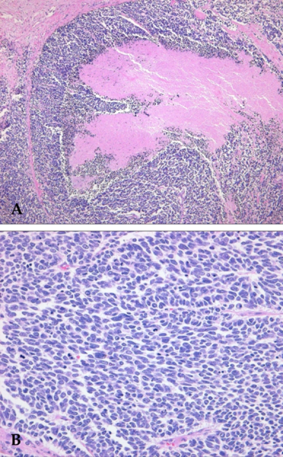
Microscopic findings of the tumor reveal solid small round cells and necrosis (A). The tumor cells show oval to fusiform hyperchromatic nuclei and indistinct nucleoli with frequent mitoses (B).

Microscopic findings of the tumor reveal solid small round cells and necrosis (A). The tumor cells show oval to fusiform hyperchromatic nuclei and indistinct nucleoli with frequent mitoses (B).