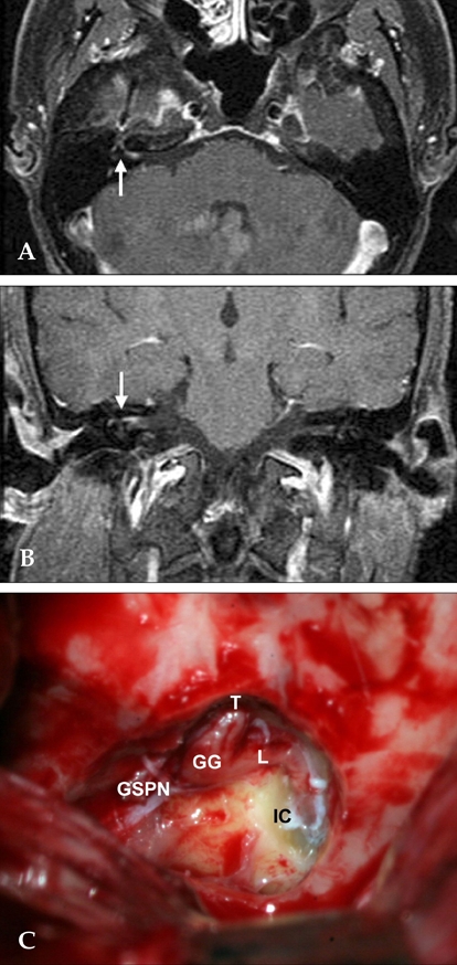
Fig. 1.

MRI and operative findings in a patient with Ramsay Hunt syndrome (right). (A) Axial contrast enhanced T1 image demonstrates enhancement from the lateral canalicular to the proximal tympanic segment of right facial nerve (arrow). (B) Coronal T1 weighted Gd-MRI shows enhancement of right facial nerve (arrow). (C) Photograph shows edematous swelling along the decompressed segment of facial nerve in MRI enhanced areas. IC, intracanalicular segment; L, labyrinthine segment; GG, geniculate ganglion; T, tympanic segment; GSPN, greater superficial petrosal nerve.
