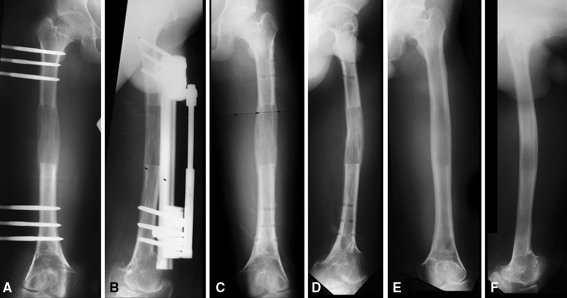
Fig. 2A–F.
The following are radiographs of a patient showing no cortices at the time of fixator removal. These include anteroposterior (A) and lateral (B) radiographs taken immediately before removal of the fixator, anteroposterior (C) and lateral (D) radiographs taken immediately following removal of the fixator, and anteroposterior (E) and lateral (F) radiographs taken 12 months after removal of fixator showing no cortices at the time of removal of the fixator and full recorticalization of the regenerate at followup without plastic deformity or fracture through the lengthened segment.

