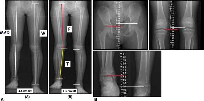
Fig. 5A–B.
(A) Standing AP radiograph of the lower extremity (modified teleoroentgenogram) performed using computed radiography on a young child with a congenital shortening of the tibia of approximately 4.5 cm. This radiograph is made with the child standing on a appropriate height lift under the short leg to level the pelvis. Besides assessing leg length discrepancy, along with length of the whole leg (W) as well as femur (F) and tibia (T), this imaging modality can be used to measure mechanical axis deviation (MAD) and joint orientation angles around the knee. (B) The modified scanogram of the same child as shown in A performed using computed radiography. Unlike a teleoroentgenogram, this imaging modality requires three radiographic exposures; one each centered over the hip, knee and ankle joints. Although a scanogram has less magnification error compared to a teleoroentgenogram, the scanogram is performed supine, is typically associated with greater radiation exposure, does not allow visualization of the entire length of the femur (F) and tibia (T) and fails to account for any shortening related to the foot. Reprinted with permission from the Journal of Bone and Joint Surgery, Inc., from Sabharwal S, Zhao C, McKeon JJ, McClemens E, Edgar M, Behrens F. Computed radiographic measurement of limb-length discrepancy. Full-length standing anteroposterior radiograph compared with scanogram. J Bone Joint Surg Am. 2006;88:2243–2251 [38].

