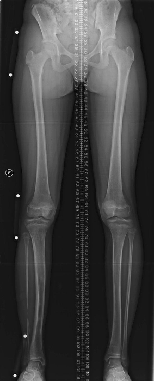
Fig. 7.

A standing full-length computed radiograph (modified teleoroentgenogram) of a 14 year old patient following right sided tibial lengthening for a 6 cm LLD. Note the use of a midline ruler and magnification markers adjacent to the right hip, knee and ankle joints to decrease the magnification error in measuring the residual LLD in this child. Use of a small lift under the right leg to level the pelvis may also have been useful.
