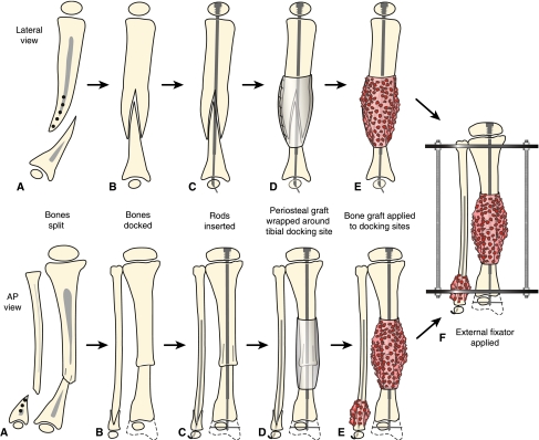
Fig. 2A–F.
Treatment for Type I CPT in the lateral and anteroposterior (AP) views is shown. (A) There is longitudinal splitting of the proximal tibial and fibular fragments. (B) The bone ends are docked and (C) IM rods are inserted (Fassier-Duval telescopic IM nail with Paley modification illustrated) into the fibula and tibia. (D) Periosteal graft is wrapped around the pseudarthrosis and (E) iliac crest bone graft is applied to the tibial and fibular docking site. (F) External fixator is applied.

