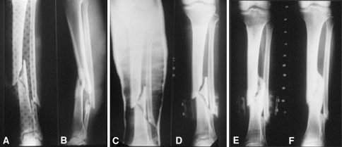
Fig. 2A–F.
(A) Anteriorposterior and (B) lateral radiographs show comminuted fractures of the tibia and fibula with severe recurvatum deformity but acceptable shortening. Radiographs were obtained after application of the original above-knee cast (C) and functional brace (D), respectively, with no attempts having been made to regain length. (E) Healing at the time of brace removal shows complete radiographic healing. (F) At final follow up solid radiographic healing is evident.

