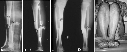
Fig. 7A–E.
(A) The radiograph shows high energy-produced comminuted fractures of the tibia and fibula. A free fragment is evident posteriorly. The shortening was accepted because the patient was elderly and had diabetes. (B) This radiograph shows the fracture as seen through the brace. (C) The anterioposterior radiograph shows the healed fractures without additional shortening or angulation, also evident (D) in the lateral view. (E) The photograph of the patient’s leg shows mild shortening of the fractured leg, which was compensated with a shoe lift.

