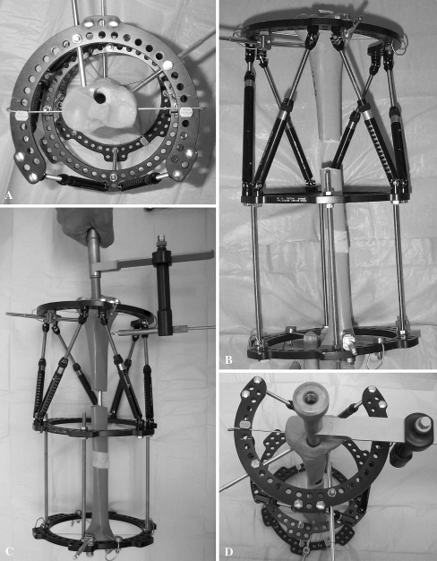
Fig. 1A–D.
This is a sawbone model illustrating LATN technique. (A) An axial view shows proximal fixation placed peripherally to avoid contact with future intramedullary nail. (B) This is a front view after 4-cm lengthening; note absent fixation on the middle ring. (C) Insertion of intramedullary nail is shown; note the targeting jig (EBI, Biomet Trauma, Parsippany, NJ) is not blocked by frame. (D) A Front axial view shows the proximal pin configuration and that the intramedullary nail is not blocked by the external fixation.

