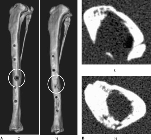
Fig. 4A–B.
(A) A reconstruction image of the whole tibia at postoperative Week 8 shows complete bridging in the human hepatocyte growth factor (hHGF) group (H), but not in the control vector group (C) with a partial defect in cortical bone. (B) An axial image at the gap level 8 weeks after the operation shows thick, circular cortical bone in the hHGF group (H), but not in the control vector group (C) with a partial defect of cortical bone.

