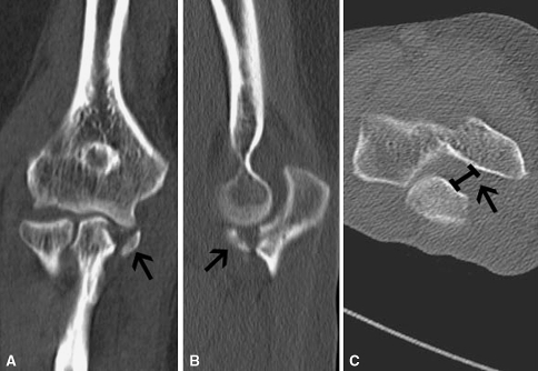
Fig. 1A–C.
(A) Anteroposterior and (B) lateral CT images show an isolated Regan and Morrey Type II coronoid fracture. (C) The axial-cut CT image at the level of the ulnohumeral joint shows considerable posteromedial instability that is not appreciated on routine examination or radiographs. Also visible is posteromedial widening or the sag sign (arrow) of the ulnohumeral joint indicative of posteromedial rotary instability.

