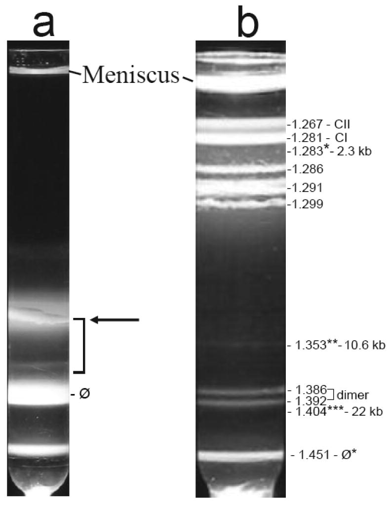
Figure 2.

Preparation of ipDNA-capsids. Cesium chloride density gradient centrifugation in two stages was used to prepare ipDNA-capsids, as described in the Materials and Methods section. (a) Light scattering is shown from the step gradient. The arrow points to a diffuse band primarily from a mixture of capsid I, capsid II and fragments of host cell. (b) Light scattering is shown from the buoyant density gradient used to further fractionate the particles in the bracketed region of the gradient in (a). The symbol, ϕ*, indicates a band formed by bacteriophage particles. The symbol, CI, indicates capsid I; the symbol, CII, indicates capsid II. The three ipDNA-capsid fractions analyzed by cryo-EM are labeled with *, ** and *** and their corresponding density and ipDNA length.
