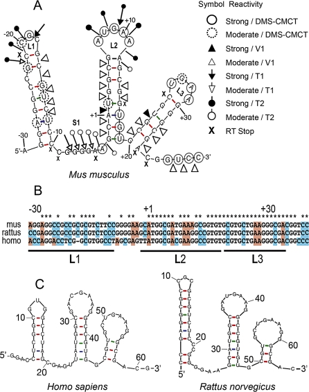
Figure 3. Secondary Structure of the SoSLIP RNA Fragment.
(A) RNA secondary structure model of the mouse Sod1–64 RNA fragment (SoSLIP) showing results from enzymatic cleavage and chemical modification experiments. White and black arrows represent moderate and strong RNase T1 cleavage sites, respectively. White and black triangles represent moderate and strong RNase V1 cleavage sites, respectively. Symbols used to indicate the reactivity of different drugs or nucleases are shown in the figure; “X” represents RT pauses.
(B) Alignment of SoSLIP sequence in mouse, rat, and human.
(C) Conservation of the SoSLIP RNA secondary structure in rat and human.

