
Fig. 6.
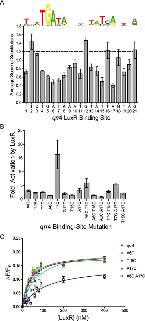
LuxR binds and regulates qrr4.
A. The consensus PWM for the LuxR binding site is shown for comparison with the actual binding site in the qrr4 promoter. The wild-type binding site is scored at 1.2 (dashed line). The average SVM score for the three substitutions at each base is presented versus the location in the binding site. Error bars indicate the standard deviation of the mean score for mutations at each position.
B. In vivo activation of qrr4–gfp expression by LuxR is shown for the wild-type promoter, single- and double-point mutants. Fold activation was calculated as the ratio LuxR+/LuxR-. Error bars represent the standard deviation of the mean ratio from two independent experiments.
C. DNA binding curves for LuxR binding to the wild-type qrr4 binding site (red), qrr4 A6C (light blue), qrr4 T15C (green), qrr4 A17C (dark blue) and qrr4 A6C, A17C (purple). The fractional change in anisotropy is plotted against the concentration of LuxR (nM). Error bars represent the standard deviation of three measurements.