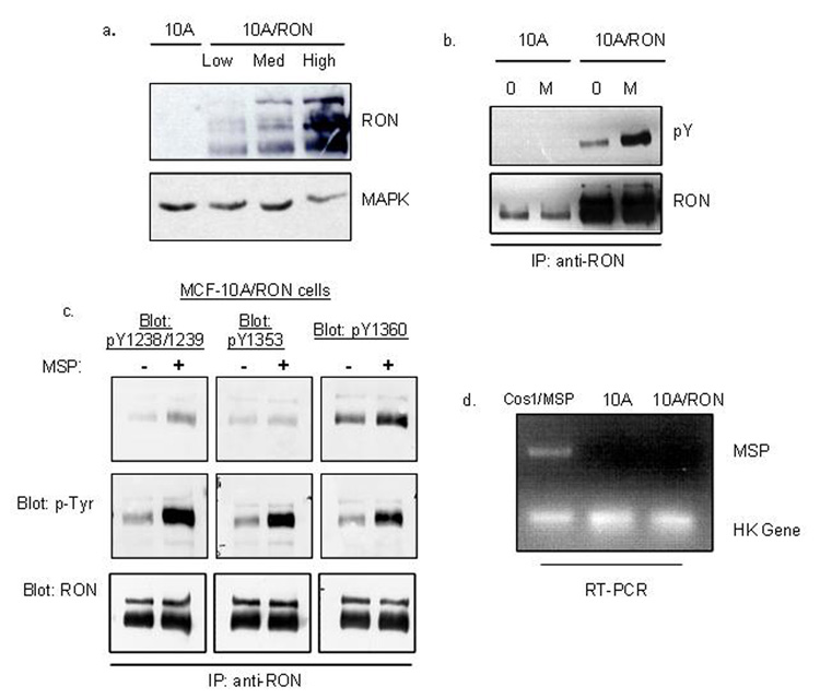
Figure 1. RON exhibits MSP-independent tyrosine phosphorylation in 10A cells.
(a) GFP-positive cells were selected by FACS analysis to yield a 10A/Vector control cell line and RON-expressing cells. RON-expression levels were detected by Western blot with an anti-RON antibody. This antibody recognizes the three different processed forms of RON: the 170 kDa unprocessed form, the 150 kDa processed and phosphorylated form and the 145 kDa processed and unphosphorylated form. Total MAPK level was used as a loading control. (b) 10A/Vector and 10A/RON cells were immunoprecipitated with an anti-RON antibody and subjected to a Western blot analysis for phospho-tyrosine (4G10 antibody). The blot was reprobed for total RON expression. (c)The same experiment as in (b), blotting with antibodies against catalytically activated RON (pY1238/Y1239), and specific tyrosine phosphorylated docking sites in the C-terminus of RON (pY1353, pY1360). (d) Semi-quantitative RT-PCR detected MSP mRNA levels. COS1 cells were transfected with a vector containing an MSP cassette as a positive control. These experiments were repeated at least three times, and shown here are representative images.

