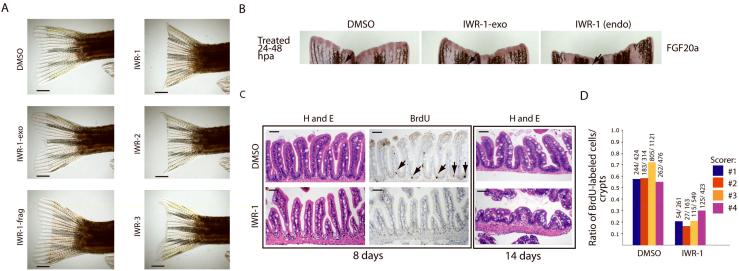
Figure 5. Chemical inhibition of the Wnt/β-catenin pathway in regeneration of zebrafish tissue.
(a) Specific inhibition of tailfin regeneration with IWR compounds. IWR-1, -2, and -3 but not the inactive IWR-1-exo and IWR-frag inhibit tailfin regeneration. Four animals in each group were analyzed. Scale bar: 2.5mm. (b) IWR-1 inhibits FGF20A expression in resected tailfins. Fish with resected tailfins were allowed to recover for 24hrs and then treated with small molecules for an additional 24hrs post amputation (hpa). Fins were probed for FGF20A expression by in situ hybridization. Arrows highlight a region that more readily reveals differences in FGF20a expression. (c) IWR-1 blocks normal homeostatic renewal of the GI tract. Representative histological sections of mid-intestinal tissue from fish treated with carrier or IWR-1 (10μM) for 8 or 14 days stained either with hematoxylin and eosin (H&E) or for BrdU incorporation. Loss of BrdU-labeled cells in the base of intestinal folds in IWR-1-treated fish (8 days; arrows) is followed by gross changes in intestinal tissue architecture after prolonged chemical exposure (14 days). Eight animals in each group were analyzed. Scale bar: 50μM. (d) Quantification of BrdU-labeled cells in the intestinal tract of control or IWR-1-treated fish. Histological sections as seen in b (middle column) were scored for the percentage of intestinal folds that contain BrdU-labeled cells. Four independent scorers analyzed sections from eight fish either from control or IWR-1 treated groups. Ratio: BrdU-labeled cells in the numerator and the number of intestinal folds scored in the denominator.

