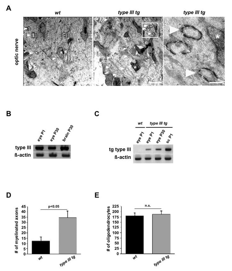
Figure 8. NRG1 type III stimulates premature myelination of the optic nerve.
(A) Electron micrographs of developing optic nerves from wildtype (left) and Nrg1 type III transgenic mice (middle) at age P6, revealing single myelin profiles (arrowheads, quantified in D). Myelinated axons in boxed area (middle panel) are magnified on the right. Asterisks mark nuclei of oligodendrocytes. Scale bars, 5 µm.
(B) By RT-PCR, Nrg1 type III mRNA is detectable in the eye of wildtype mice at age P1 and P30 (brain mRNA serving as internal positive controls; β-actin internal control).
(C) By transgene-specific RT-PCR, type III mRNA is detectable in the eyes of postnatal (P1 and P30) Nrg1 type III trangenic mice, but not in wildtype (wt). Spinal cord mRNA (sc, age P1) as positive internal control; β-actin internal control.
(D, E) Higher number of myelinated axonal profiles (in D; p<0.05) but equal number of oligodendrocytes (in E) in the optic nerve of 6 days old Nrg1 type III transgenic mice (grey bars), when compared to age-matched controls (black bars). Quantification is from semithin cross sections (wt, n=5; Nrg1 type III transgenics, n=6). Error bars, SEM (unpaired, two-sided t-test).

