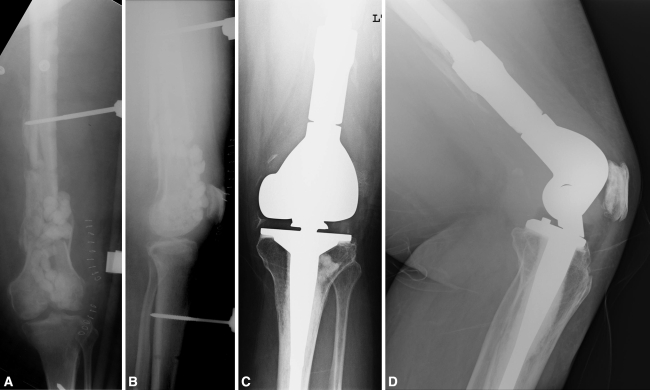
Fig. 2A–D.
A 52-year-old woman sustained a high-energy distal femur fracture initially treated with open reduction and internal fixation. This went on to become an infected nonunion. After hardware removal and placement of an external fixator and antibiotic-laden beads, the patient presented for evaluation. (A) Anteroposterior and (B) lateral radiographs show a comminuted nonunion with an external fixator and antibiotic beads in place. (C) Anteroposterior and (D) lateral radiographs show the large segmental distal femoral replacement device used during reimplantation after two-stage radical débridement and an intervening period of antibiotic therapy.

