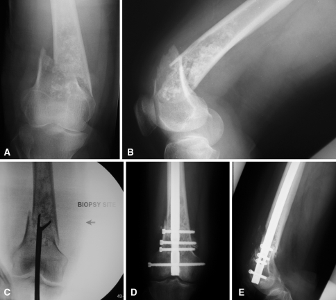
Fig. 1A–E.
Preoperative (A) anteroposterior and (B) lateral radiographs of a 65-year-old woman with a pathologic fracture of the right distal femur show a large intramedullary lesion with ill-defined borders, diffuse endosteal scalloping, and cartilaginous matrix production at the level of the fracture. (C) An intraoperative fluoroscopic image shows a joint-violating intramedullary biopsy of the chondrosarcoma. Postoperative (D) anteroposterior and (E) lateral radiographs show stabilization of the fracture with a retrograde intramedullary nail, violating the chondrosarcoma and contaminating the proximal femoral canal.

