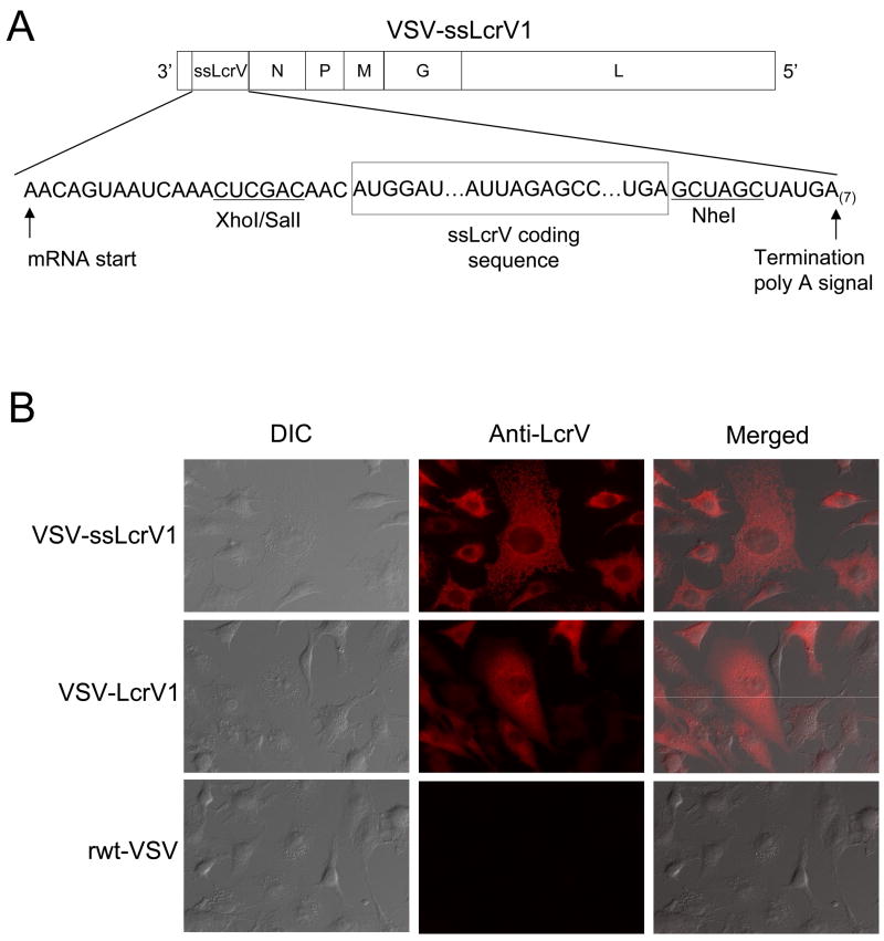
Figure 1.
Recombinant VSV expressing the secreted form of Yernisia pestis lcrV gene (A) Schematic representation of the recombinant VSV with ssLcrV inserted upstream of the N gene (VSV-ssLcrV1) used in this study, showing the gene order in 3′ to 5′ direction on the negative-strand genome. (B) Indirect immunofluorescence microscopy of BHK-21 cells infected with the indicated viruses. Cells were fixed at 3 hrs post infection, permeabilized and stained using anti-LcrV monoclonal antibodies (MAbs), followed by an AlexaFlour-594 secondary antibody. The differential interference contrast (DIC) images are shown in the left column, the fluorescence images are shown in the middle column and the merged images are shown in the right column.

