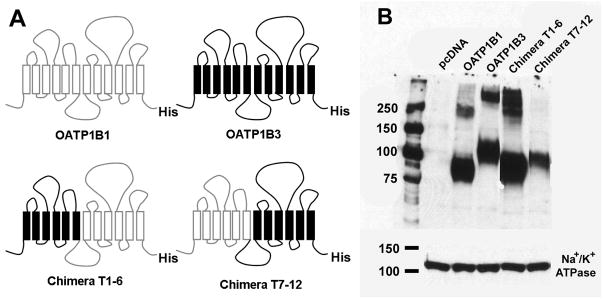
FIGURE 1.
Schematic representation of human OATP1B1, 1B3 and chimeras T1–6 and T7–12, and their surface expression in HEK293 cells. (A) Chimera T1–6 consisted of TMs 1–6 (residues 1–275) of OATP1B3 and TMs 7–12 (residues 276–691) of OATP1B1, while chimera T7–12 consisted of TMs 1–6 (residues 1–275) of OATP1B1 and TMs 7–12 (residues 276–702) of OATP1B3. In order to detect all constructs with the same antibody, a His-tag was introduced at the C-terminal end of all constructs. (B) Western blot analysis of surface biotinylated proteins detected with an anti-His antibody. The plasma membrane marker Na+/K+ ATPase a-subunit was used as protein loading control.

