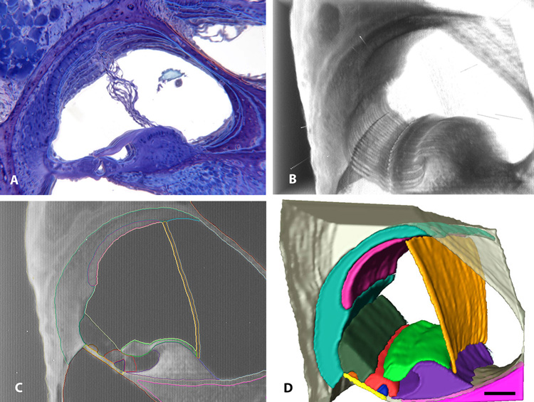
Fig. 5.
A four-panel figure showing serial sections of a mouse cochlea from celloidin sections (A), and OPFOS sections in which segmentation and 3D reconstruction of cochlear structures was performed. (A movie of the OPFOS sections is also available in the Supplementary Materials of this paper and on the MCD website). A. Amira alignment and registration of 13 images from the celloidin serial sections which show poor alignment of cochlear structures due to mechanical sectioning artifacts of the tissue. B. Direct volume rendering of 24 OPFOS images shows excellent alignment of the sections using Amira. C. At least 14 different cochlear structures could be outlined (i.e., segmented) using different colors. D. Eleven of the 14 different structures were prepared as 3D isosurface renderings. Bar = 100 µm in all four panels.

