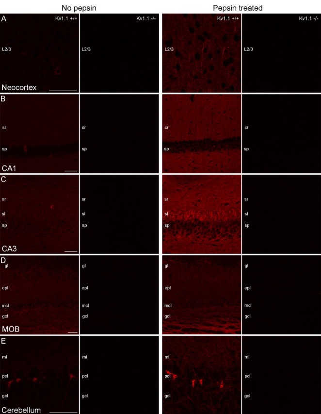
Figure 1.

Testing the specificity of Kv1.1 subunit immunolabeling on conventional and pepsin-treated brain sections using Kv1.1−/− mice. A–D, Without pepsin treatment, a faint neuropil or cytoplasmic labeling in scattered cells is detected in the neocortex (A, left), CA1 (B, left), and CA3 (C, left) areas of the hippocampus and in the MOB (D, left) obtained from Kv1.1+/+ mice, and all labeling disappears in Kv1.1−/− mice. After pepsin treatment, the cytoplasmic labeling of scattered nerve cells is not observed, but the neuropil labeling became more pronounced and intensely labeled AISs appear (A–D, right). In Kv1.1−/− mice, all immunosignals disappear. E, In the cerebellar cortex, the pinceau is strongly immunopositive for the Kv1.1 subunit with and without pepsin treatment. The specificity of immunosignal is proven by the lack of labeling in Kv1.1−/− mice. Images from Kv1.1+/+ and Kv1.1−/− sections were acquired with identical settings (e.g., PMT voltage, laser intensity) of the confocal microscope. Within each panel, all images are at the same magnification. Scale bars, 50 μm.
