Figure 5.

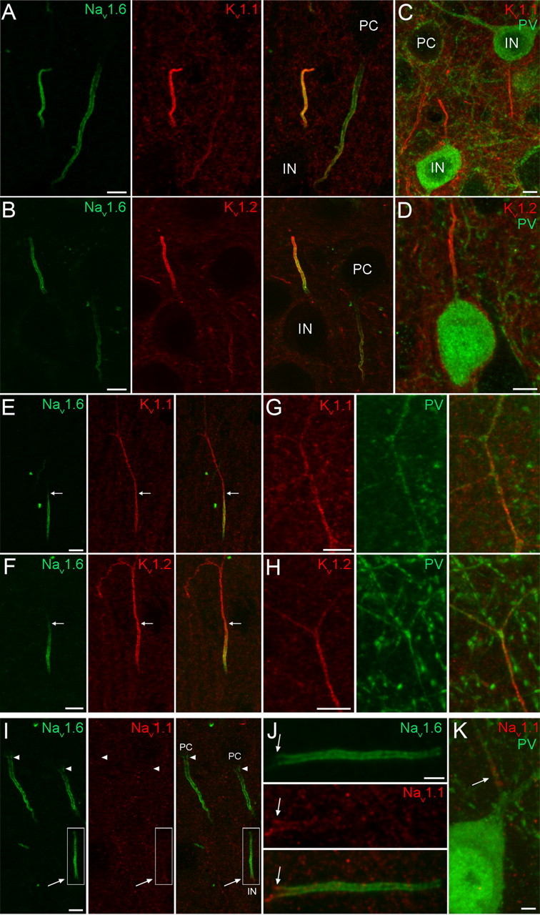
Localization of the Nav1.1, Nav1.6, Kv1.1, and Kv1.2 subunits in the AIS of neocortical INs. A, B, Double immunofluorescent reactions showing the colocalization of the Nav1.6 subunit with either Kv1.1 (A) or Kv1.2 (B) subunits in neocortical IN AISs. The AISs of GABAergic INs are more intensely labeled compared with the neighboring PC AIS. C, D, Strongly Kv1.1 (C) and Kv1.2 (D) immunoreactive AISs emerge from PV immunopositive INs. E, F, In a subpopulation of INs, in contrast to the Nav1.6 subunit labeling, the Kv1.1 (E) and Kv1.2 (F) labeling is not restricted to the AIS, but extends into the ascending axon. Arrows demarcate the border of the AIS and more distal axon. G, H, Some Kv1.1 (G) and Kv1.2 (H) subunit immunoreactive axons are also immunopositive for PV. I, Double immunofluorescent labeling reveals the absence of Nav1.1 subunit immunoreactivity in PC AISs (arrowheads) and its presence in an IN AIS (arrow) emerging from the apical pole of the IN soma. J, A higher-magnification view of the boxed area in I shows that the Nav1.1 subunit immunolabeling is confined to the proximal part (arrow) of the AIS. K, Nav1.1 subunit immunolabeling is present in the proximal part of an axon emerging from a PV immunopositive cell. Scale bars: A–I, 5 μm; J, K, 2 μm.
